A biotech growing engineered muscle tissue
Count every fiber. Skip the day at the microscope.
A day at the microscope becomes minutes, and the number matches the human expert: within ±10% on 85% of images, across 119 validated images.
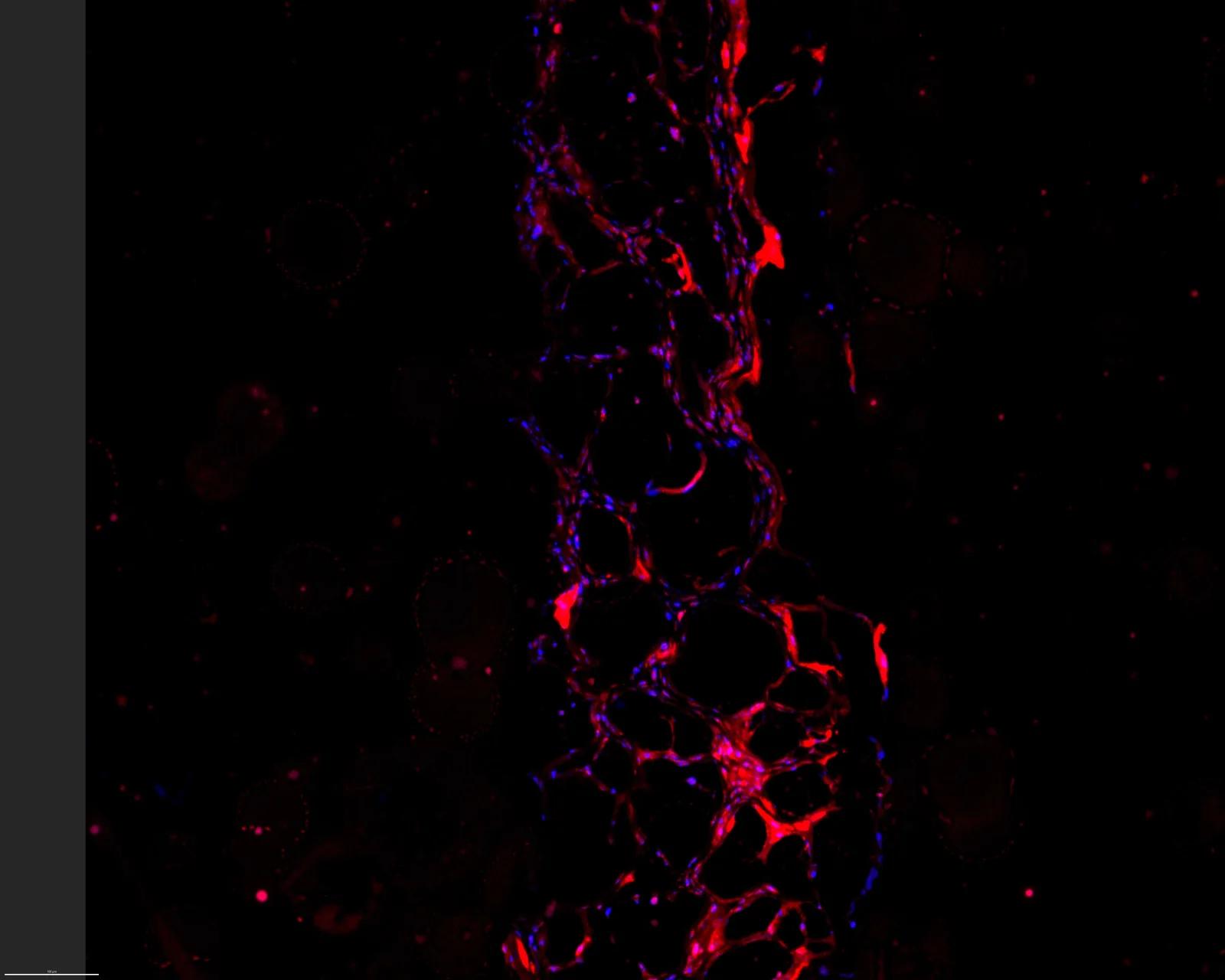
The number the expert would have written down, in 1% of the time.
THE COST
Measuring fibers by hand eats days
Counting and measuring muscle fibers in a microscopy image is slow expert work. In Fiji or ImageJ, a person marks hundreds of fibers per image, by hand, over and over. One image takes hours. A full set takes days.
And it isn't only time. Two people count the same image and get two numbers. The team growing the tissue waits on that number to decide what happens next.
NO BLACK BOX
You can see how every number was reached
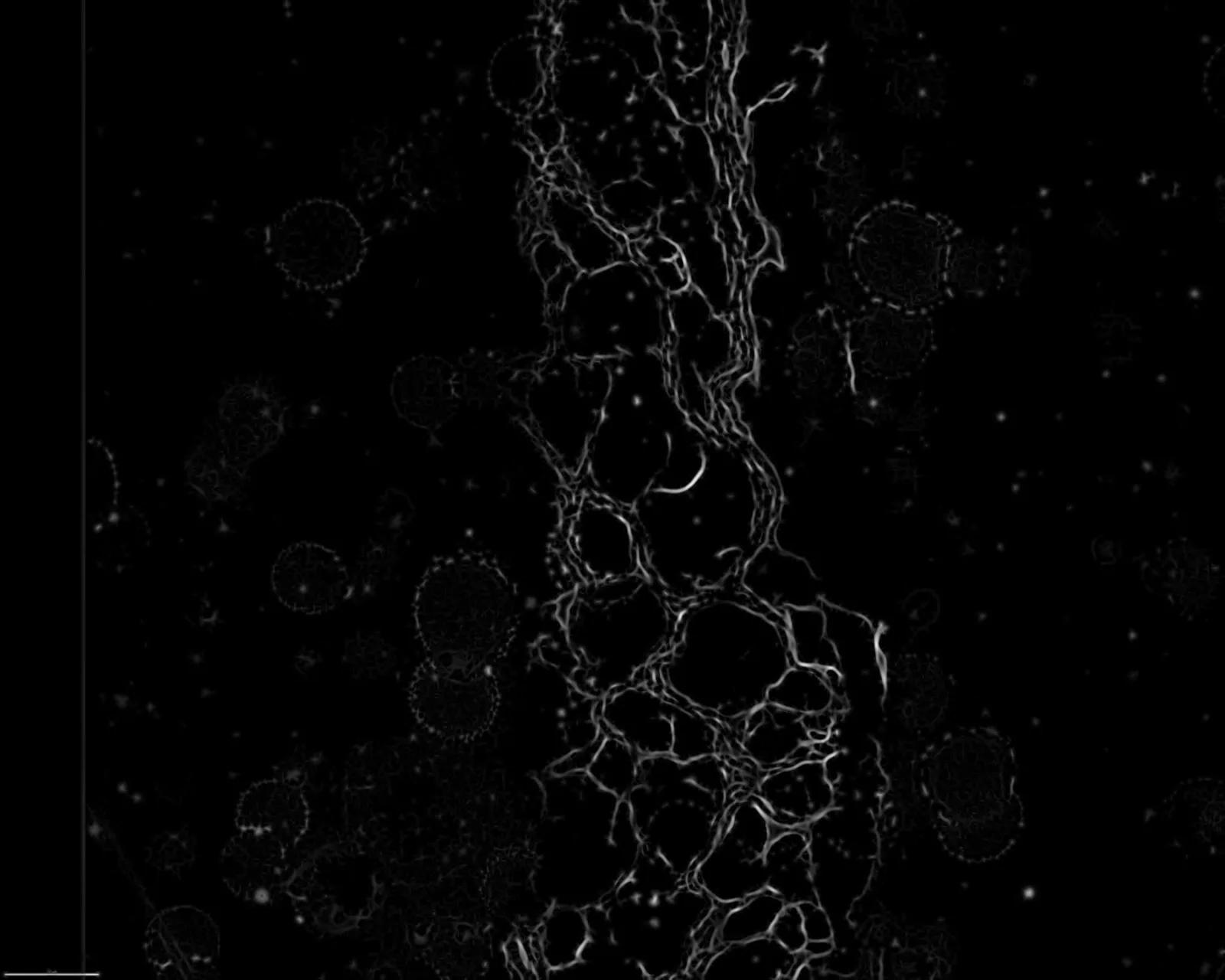
Trace each wall
It traces the wall of every fiber in the image.
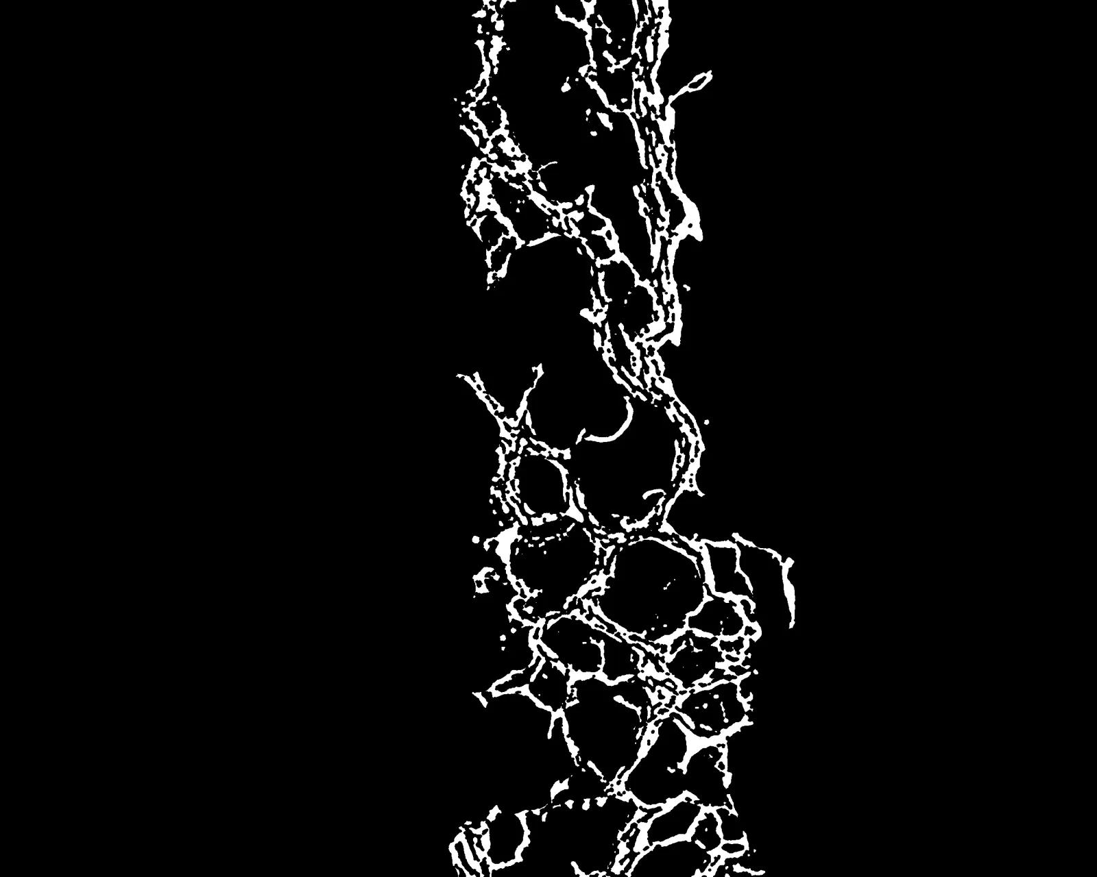
Separate touching fibers
Fibers that touch are separated, so each one is counted once.
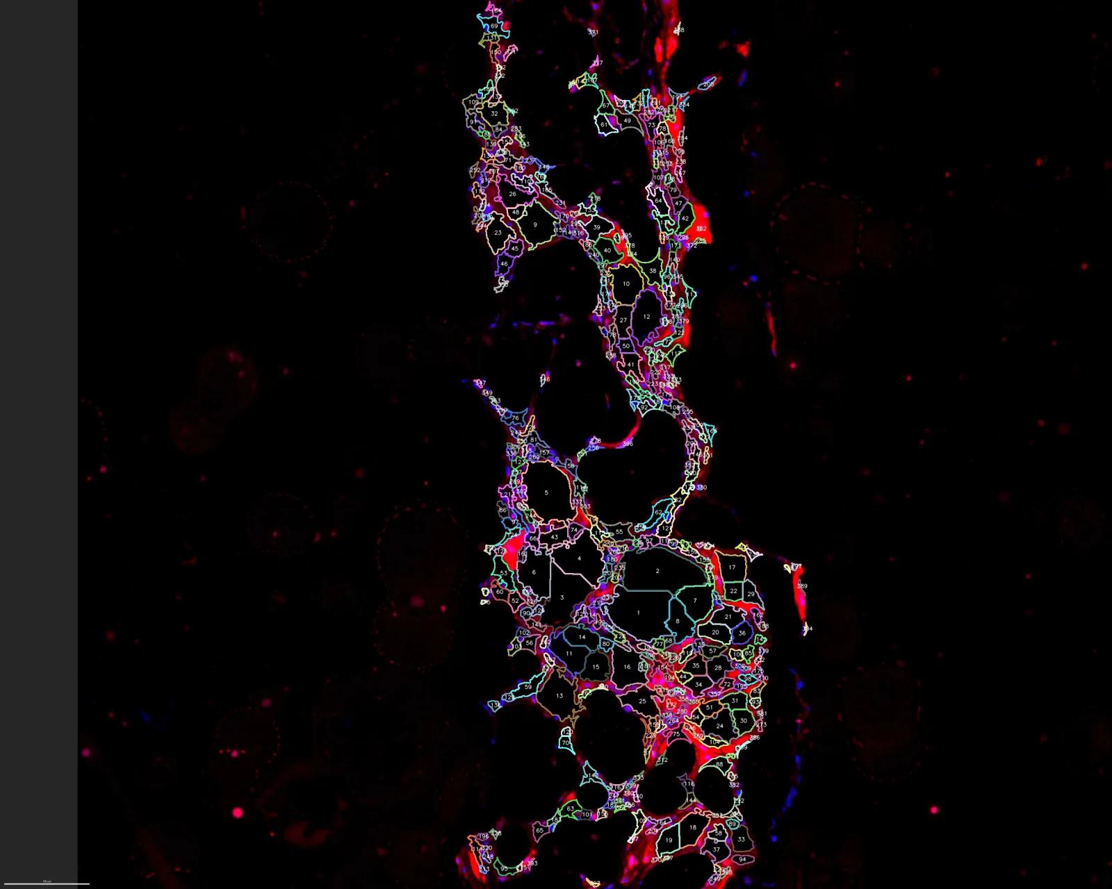
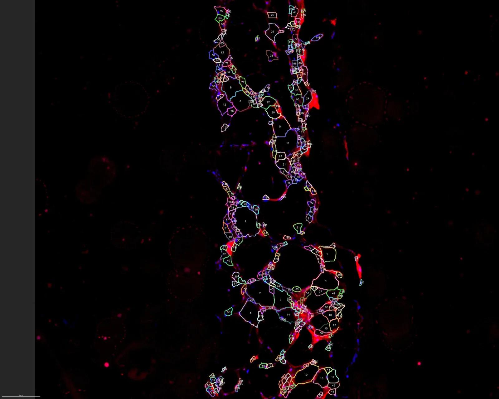
Count and measure
Every fiber is outlined, counted and measured, with a check layer you can review.
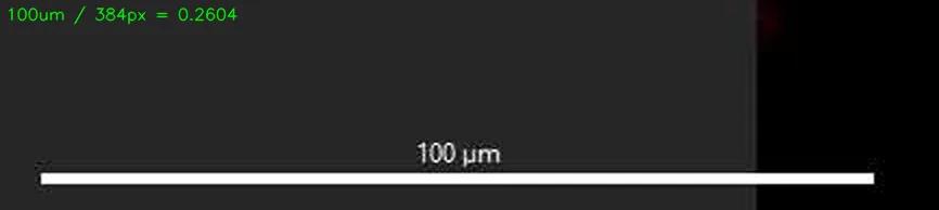
It reads the scale bar automatically to convert pixels to microns. No manual input, no run-to-run drift. Same image, same number, every version, and every step logged.
THE RESULT
Not 'close enough.' The expert's number.
Across 119 images from two biopsies, the median measurement sat within ~0.7% of the manual count, and 85% within ±10%. Not "close enough." The number the expert would have written down.
Validated on two biopsies across 119 images. We re-validate on your tissue before you rely on it.
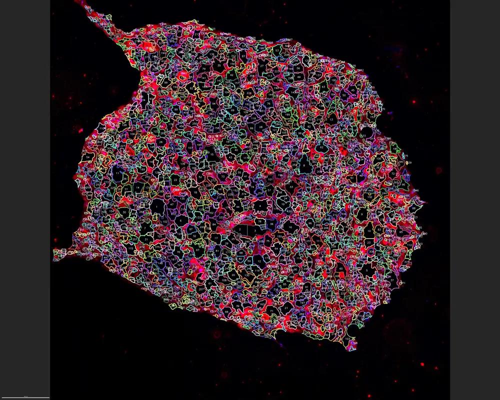
Even where fibers crowd, each is counted once.
Coming next
Cross-sectional area (CSA), in clinical validation. The overlays show the method; the accuracy figures aren't final.
Have microscopy work eating your team's days?
Let's see if this fits your images.
Book a 20-min fit check