A biotech growing engineered muscle tissue
Minutes an image became seconds. Ten at a time.
Two slow, manual stages (preparing the mCherry channel and measuring the fibers) now run automatically: from minutes an image to seconds, ten images at once, every parameter under the scientist's control.
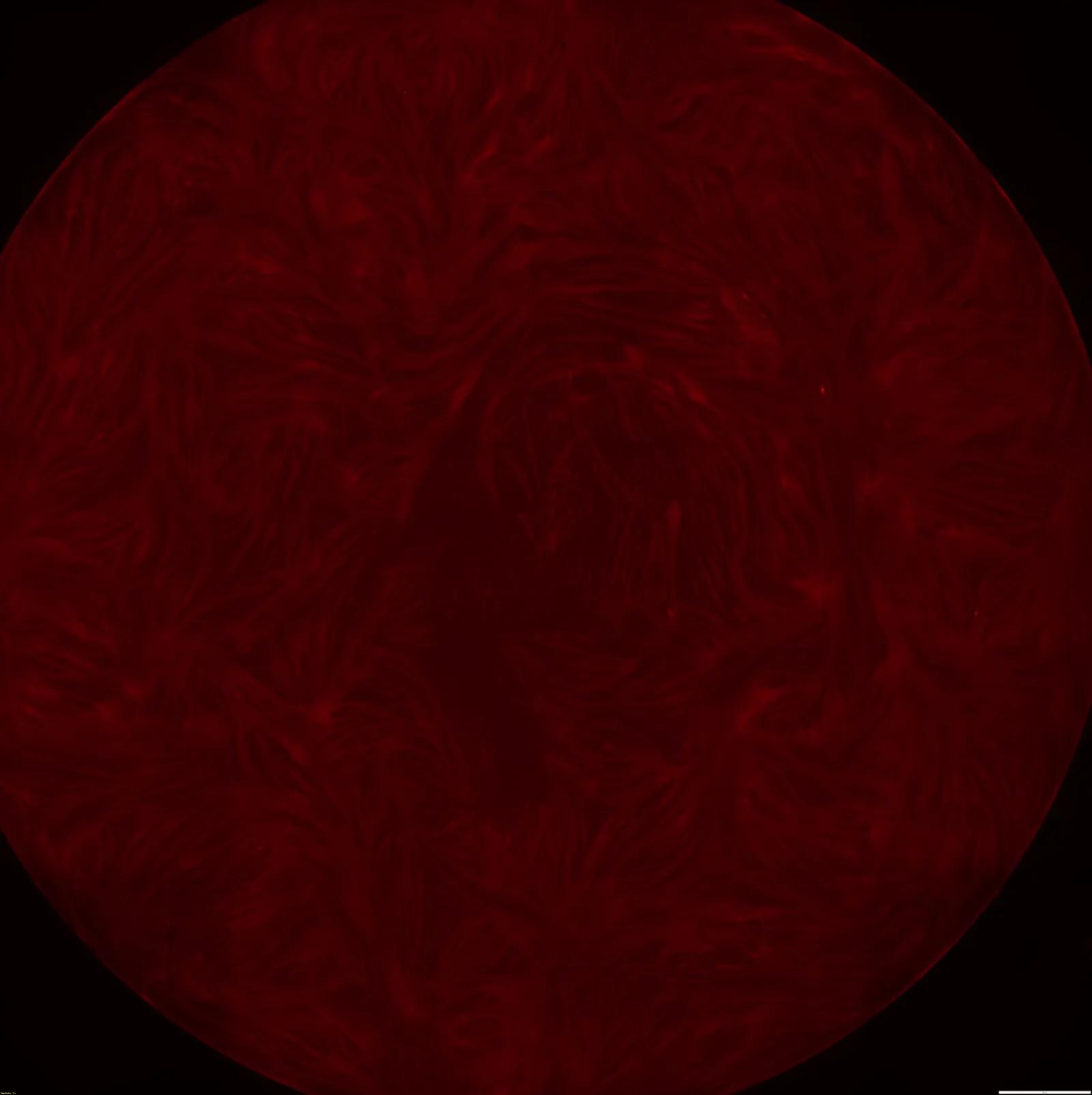
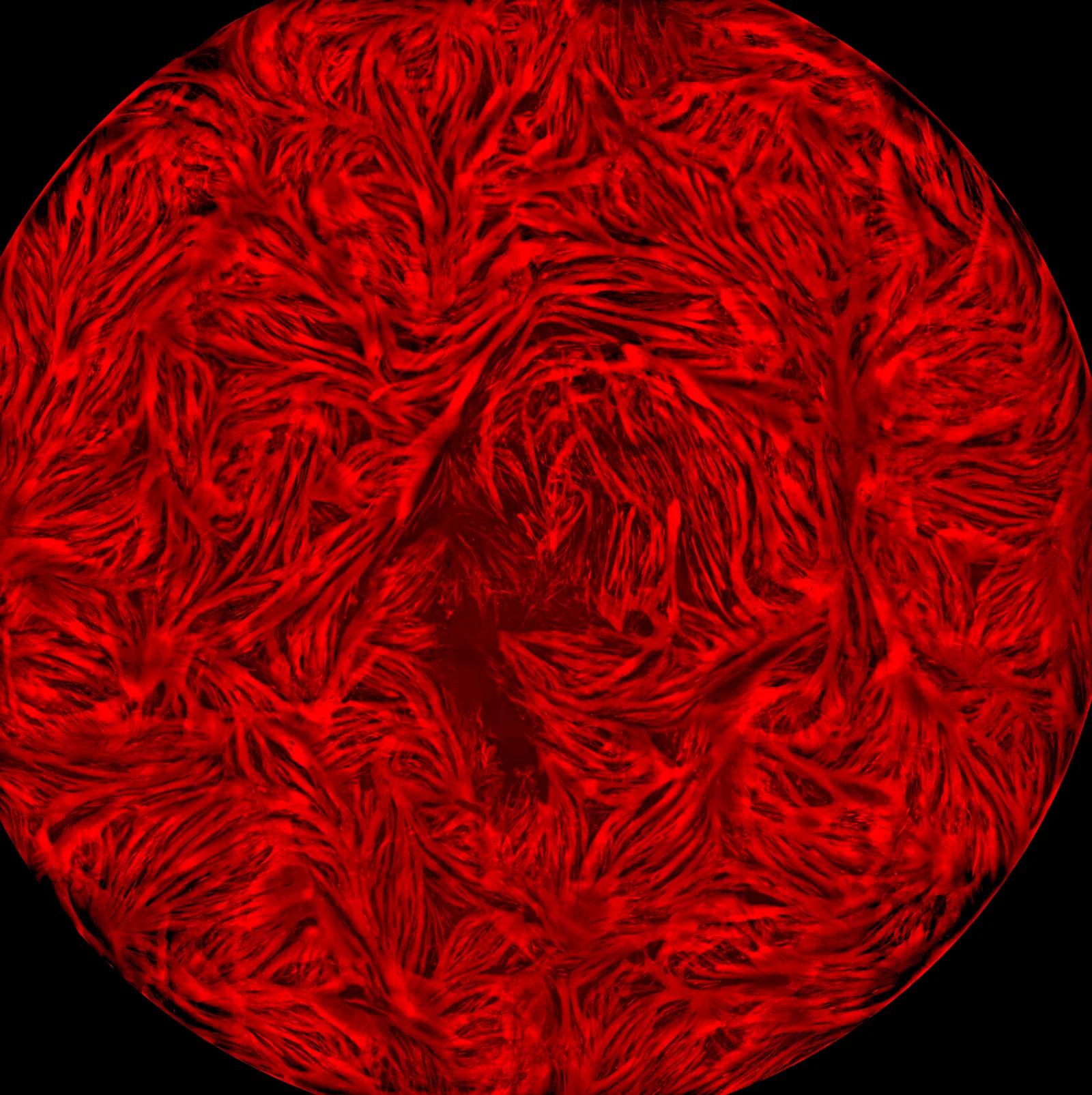
5–8 minutes of hand-work per image, now 3–5 seconds.
Two manual stages that ate minutes per image (prep and measurement) now run in seconds, ten images at once.
THE COST
Two manual stages, before anyone even measured
Before any measurement, a person prepared every image by hand. The mCherry channel comes off the microscope dim, and getting it measurement-ready meant a Fiji macro, then manual Levels and Curves tuning in Photoshop until the fibers popped. 5–8 minutes an image, hundreds of images a month, and every operator a little different.
Then came the measurement script, a black box another company had written. It was slow, 2–3 minutes an image, ran one image at a time with no multithreading, and let the research team change nothing, not even one parameter. Slow, and out of their hands.
THE MEASUREMENT
Every fiber measured and marked on the image itself
We rebuilt the measurement. It runs on the prepared mCherry channel, measures the width of every fiber and marks it directly on the image: an ID and a width per fiber, visible to check. Under 20 seconds an image, and ten in parallel.
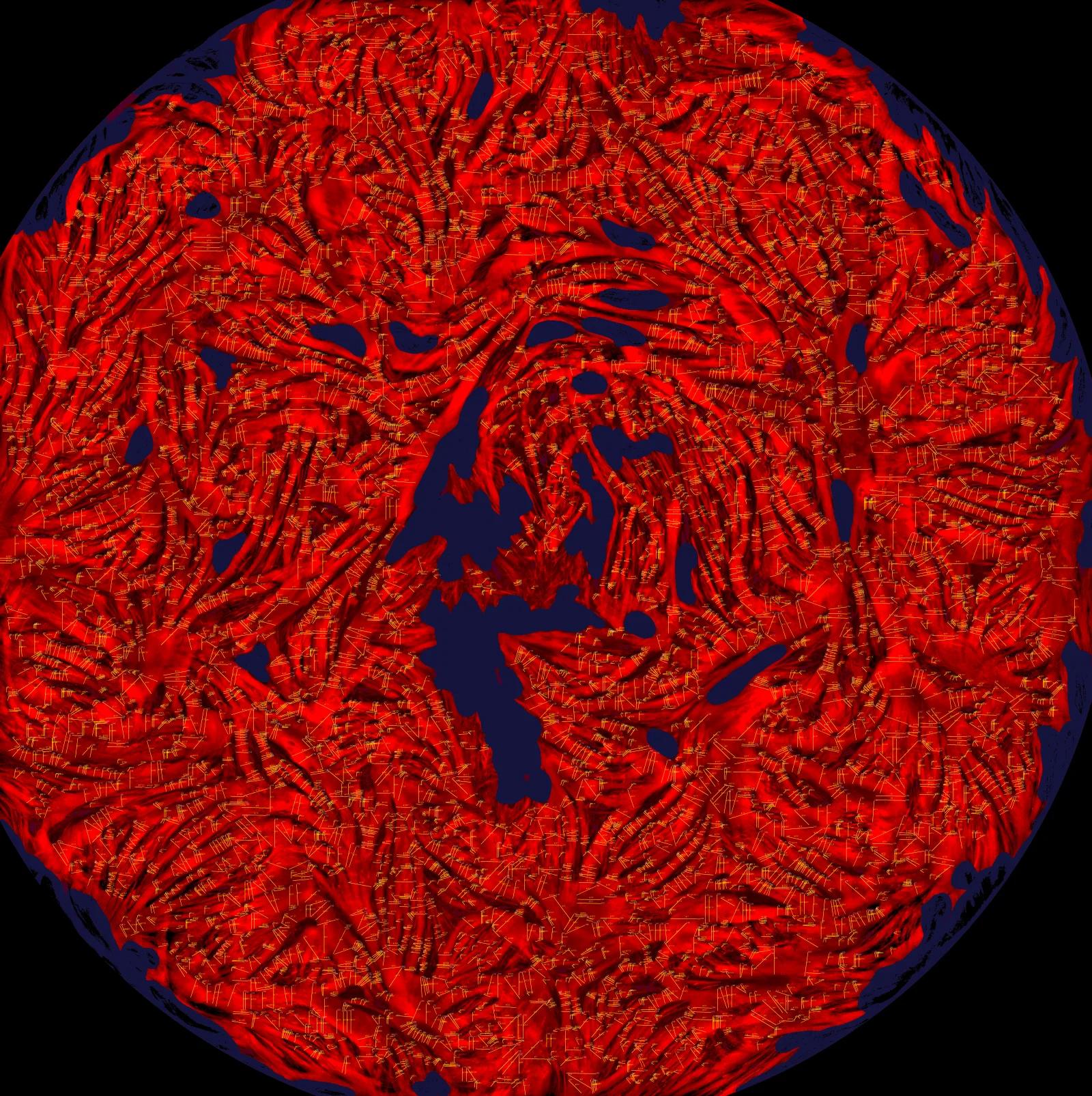
The result on the image itself: a width and an ID for every fiber, thousands of measurements in a single image.
FULL CONTROL
You set every parameter, without touching code
And most important: it's no longer a black box. Line spacing, morphology, void detection, confidence threshold, every parameter is visible and yours to set. You sweep 13 ready-made configs to find the one that fits your tissue, and every run is logged and reproducible: same image, same number, every time.
| config | what it tunes | fibers | QA |
|---|---|---|---|
| C01_baseline | v17 default, reference point | 288 | reference |
| C04_gentle_strict_border | team-preferred for HITL review | 301 | pass |
| C05_gentle_soft_void | softer void, keeps thin fibers | 312 | pass |
| C08_dense_sampling | denser spacing, more per fiber | 356 | review |
| C10_max_precision | strict + aggressive, highest confidence | 264 | pass |
The real fiber-width parameter sweep, with the same config names the team tunes.
A QA agent scores every measurement
Seven independent signals score every measurement line and write a short list of the images worth a human glance, usually just 3–8%. The rest you can run automatically.
And the numbers match the manual work
Across 119 images from two biopsies, the median stayed within ~0.7% of the manual prep, and 85% of images within ±10%. The same result, without the hand-tuning.
Running a measurement script over microscopy images, one by one?
Let's see how much time you'd get back, without giving up control.
Book a 20-min fit check